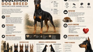
Doberman Skin Lumps and Color Dilution Alopecia Guide
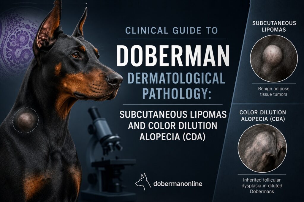
Clinical Guide to Doberman Dermatological Pathology: Subcutaneous Lipomas and Color Dilution Alopecia (CDA)
The Doberman Pinscher is a breed widely admired for its sleek, aerodynamic silhouette, aristocratic posture, and deep muscular definition. However, their unique genetic profile and specialized coat structure render them highly susceptible to specific dermatological pathologies. Discovering a sudden anatomical mass beneath the dermis or witnessing progressive alopecia (hair loss) in dilute color phenotypes can be deeply concerning for owners and breeders alike.
This clinical analysis evaluates two prominent soft tissue and genetic skin disorders frequently diagnosed in Dobermans within North American veterinary practices: Subcutaneous Lipomas and Color Dilution Alopecia (CDA).
1. Subcutaneous Lipomas: Benign Neoplastic Fatty Masses
A lipoma is a benign (non-carcinogenic) tumor composed entirely of mature adipocytes (fat cells) localized within the subcutaneous tissue overlying the musculoskeletal architecture. While they can develop at any developmental stage, they are predominantly diagnosed in senior Dobermans aged 7 years and older.
Clinical Identification and Palpation Metrics
When assessing a subcutaneous mass on a Doberman, veterinary professionals and owners evaluate the lesion based on specific physical criteria:
-
Mobility: Upon manual palpation, the mass shifts freely over and beneath the underlying anatomical structures, showing no adhesion to the abdominal wall, ribs, or muscle tissue.
-
Consistency: Elastic, soft, and localized. It feels distinctly “doughy” or rubbery and does not present the rigid, calcified, or bone-like structure associated with malignant osteosarcomas or soft tissue sarcomas.
-
Analgesic Nature: Lipomas are inherently non-painful. The canine exhibits no nociceptive (pain) response, defensive behavior, or localized inflammation/heat upon direct handling.
-
Anatomical Distribution: Most frequently observed in the axillary region (armpits), sternal border (chest), inguinal fold (groin), and proximal limbs.
+-----------------------------------------------------------------+
|                    DIAGNOSTIC WARNING BELL                      |
|                                                                 |
|  It is clinically IMPOSSIBLE to distinguish a benign lipoma     |
|  from a highly malignant Mast Cell Tumor (MCT) or soft tissue   |
|  sarcoma purely by visual observation or physical palpation.    |
+-----------------------------------------------------------------+
Etiology and Veterinary Intervention
The exact molecular origin of lipomas remains idiopathic (unknown), though genetic predisposition, metabolic slowing, and canine obesity are high-driven catalysts.
From a clinical management standpoint, because these masses are benign, surgical excision is rarely indicated unless the tumor undergoes rapid hypertrophic growth or physically restricts joint mobility (e.g., a massive axillary lipoma impeding the forelimb stride).
The Gold Standard Protocol: To secure a definitive diagnosis, a Fine-Needle Aspiration (FNA) biopsy must be performed. The veterinarian inserts a small-gauge needle into the lump to collect cells, which are then stained and examined under a microscope to confirm the presence of harmless fat droplets rather than malignant cells.
2. Color Dilution Alopecia (CDA): Genetic Follicular Dysplasia
Color Dilution Alopecia (also historically referred to as “Blue Doberman Syndrome”) is a hereditary ectodermal dysplasia that affects dogs possessing diluted coat variations. While the American Kennel Club (AKC) breed standard recognizes Black & Rust and Red & Rust as the dominant pigmentations, recessive genetic dilution mutations generate the Blue (diluted black) and Fawn/Isabella (diluted red) phenotypes.
Pathophysiology of CDA
The dilution phenotype is strictly governed by the melanophilin (MLPH) gene on the D locus. In canines affected by CDA, a defective mutation causes a chaotic distribution, structural defect, and severe clumping of melanin pigments (melanosomes) within the hair shafts and follicular units.
These resulting macromelanosomes structurally weaken the entire hair matrix. As a result:
-
The hair shafts become extremely brittle and prone to fracturing under minor friction (such as normal grooming or collar wear).
-
The hair breaks off at the skin line.
-
Over time, the repeated damage causes permanent follicular atrophy, meaning the hair follicle dies and can never grow hair again.
       [Healthy Doberman Hair]              [CDA-Affected Doberman Hair]
       +---------------------+              +--------------------------+
       | Evenly distributed  |              | Melanin Clumps           |
       | Melanin Pigments    |              | (Macromelanosomes)       |
       |                     |              |    ●●   ●●●●    ●●       |
       +---------------------+              +--------------------------+
          (Strong Matrix)                     (Brittle / Fractures Easily)
Chronological Onset and Symptomatology
-
The Juvenile Window: Affected puppies are born with seemingly flawless, lustrous, and beautiful blue or fawn coats. Clinical symptoms typically manifest during the adolescent development window, anywhere between 6 months and 2 years of age.
-
Dermatological Progression: Chronic alopecia typically begins along the dorsal midline (the spine), characterized by a dull, dry, “moth-eaten” coat appearance. Interestingly, the extremities (the head and lower limbs) are usually spared.
-
Secondary Complications: As the protective coat vanishes, the exposed epidermis becomes highly vulnerable. The skin exhibits dry scaling, flaking, hyperpigmentation (turning dark or black), and recurrent secondary bacterial folliculitis (infected, pus-filled bumps or papules caused by bacteria entering the open hair follicles).
Clinical Management Protocols for CDA
Because CDA is a fundamental genetic disorder, there is no permanent therapeutic cure to reverse the mutation or restore dead, atrophied hair follicles. Management focuses entirely on preserving epidermal barrier integrity and preventing infection:
-
Neuroendocrine Modulation (Melatonin): Oral melatonin supplementation (typically 3–6 mg, administered under veterinary guidance) can sometimes stimulate dormant follicular matrices in select patients, improving coat quality and reducing the velocity of ongoing hair loss.
-
Epidermal Barrier Restoration: Regular bathing with soothing colloidal oatmeal, moisturizing rinses, or antiseptic/keratolytic shampoos (containing sulfur or salicylic acid) is crucial to manage scaling. Furthermore, incorporating concentrated Omega-3 and Omega-6 fatty acids (high-quality marine lipid oils like wild-caught salmon oil) into their daily diet fortifies the skin’s lipid barrier from the inside out.
-
Environmental Photoprotection & Warmth: An alopecic (bald) Doberman lacks its natural shield. The exposed skin is highly vulnerable to solar radiation burns (sunburn), which can increase the risk of skin cancers, as well as winter hypothermia. Owners must apply canine-specific, non-toxic, zinc-free sunscreens during peak UV hours and utilize insulating protective garments (fleece coats or sweaters) during cold weather cycles.
FAQ: Frequently Asked Questions
Can subcutaneous lipomas in Dobermans transition into malignant carcinomas?
No. True lipomas are histologically benign and do not metastasize or undergo malignant transformation into liposarcomas (malignant fat tumors). However, Dobermans that are prone to lipomas often develop multiple lumps over their lifespan. A new lump appearing right next to an old, confirmed lipoma could easily be a completely different, malignant pathology. Therefore, every single new dermal mass should be evaluated via FNA.
Does Color Dilution Alopecia develop in all Blue or Isabella Dobermans?
Not universally, but the statistical probability is exceptionally high. Epidemiological data within North America shows that approximately 75% to 80% of Blue Dobermans, and an even higher percentage of Fawn/Isabella variations, will develop varying degrees of CDA during their lifecycle. Some lines maintain a healthier coat longer, but any dilute Doberman should be monitored closely starting at 6 months of age.
Is Color Dilution Alopecia inherently painful for the dog?
No. CDA itself is purely a structural and cosmetic disorder. The hair loss and color changes cause zero neurological pain, itching, or systemic distress to the canine. However, if the exposed, unprotected skin is left unmanaged, it will likely develop secondary deep pyoderma or bacterial folliculitis. These secondary bacterial and yeast infections cause severe pruritus (intense itching), crusting, and localized pain that absolutely requires antibiotic intervention.
Will pet insurance cover these conditions in the United States?
In the US market, most major pet insurance providers will cover the diagnostic costs (like FNA biopsies) and treatments for lipomas and secondary CDA skin infections, provided the conditions were not documented before the policy’s enrollment waiting period. Because CDA is hereditary, a few select companies may classify it under genetic/hereditary exclusions, so owners should carefully review their policy’s specific clauses regarding hereditary breed traits.